What happens to my tooth when I have to have fillings, crowns, bonding or any other restorations?

What happens to my tooth when I have to have fillings, crowns, bonding or any other restorations?


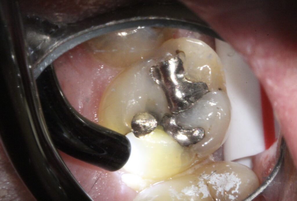
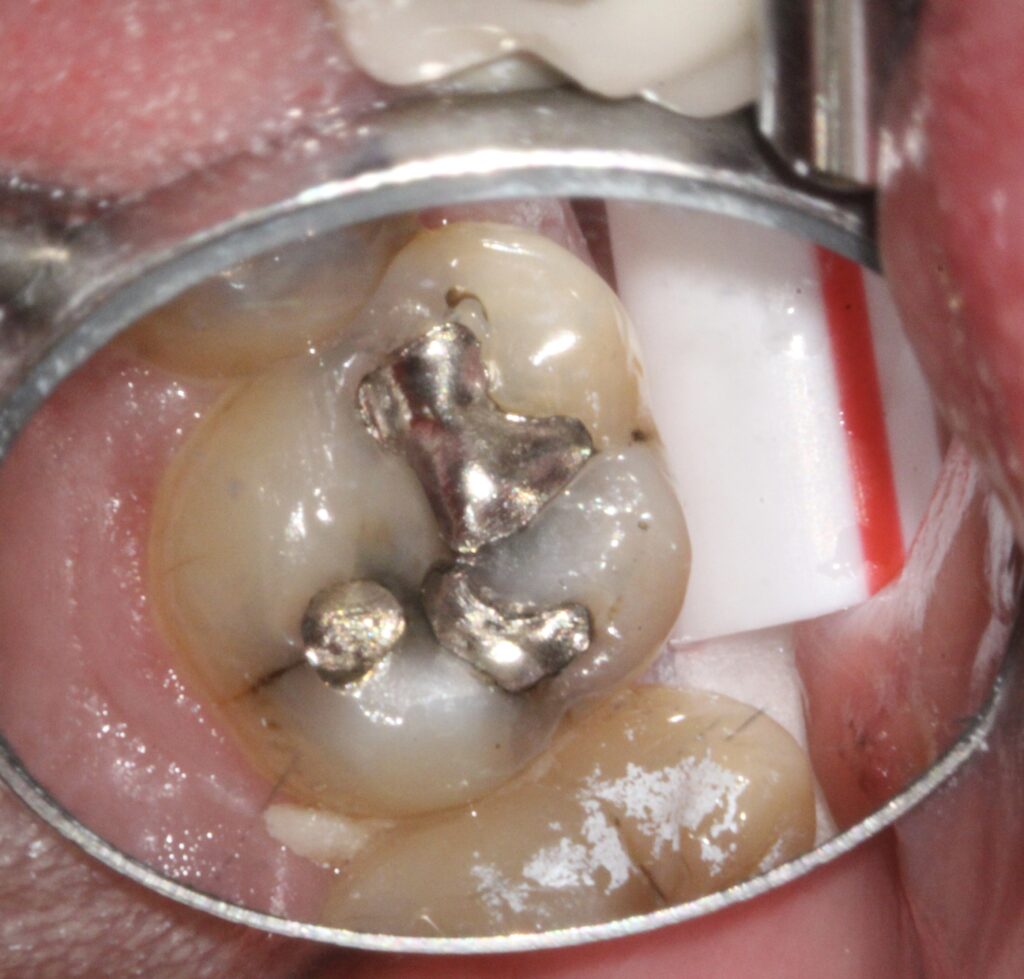
To perform dental restorations, the diseased tooth structure and all the old filling material/crowns/etc. have to be removed.  Any time a dental drill contacts a tooth, the drill’s rotation causes some tooth trauma.  Lots of water and slower drilling speeds are used to minimize trauma to the tooth’s nerve.  Unfortunately, there is no alternative to drilling on a tooth to remove old restorations (dental lasers cannot cut old fillings or crowns).  Sometimes the actual drilling on the tooth causes postoperative sensitivity and the tooth becomes more sensitive to cold.  This is usually temporary but may last for several months.


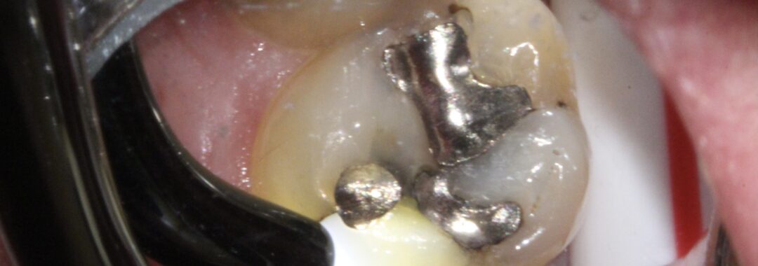
Teeth that have had a lot of fillings or crowns may also experience more sensitivity after treatment.  The difficulty with assessing the health of these teeth, is that we cannot remove them and look at their nerve tissue under a microscope without permanently removing them from patients.  Teeth with lots of dental history have had a lot of nerve trauma over their life span.  Sometimes these teeth appear to still be healthy and respond well to our nerve tests when in reality there is dead nerve tissue already present and we cannot see it inside the tooth.  These teeth may eventually demonstrate signs and symptoms of necrotic nerves and need root canals.  We always try to determine nerve health prior to restoring teeth, but sometimes it is just not possible to be accurate due the long dental history of some teeth.  That being said, we still only recommend root canals for teeth with apical periodontitis (signs and symptoms of dead nerves) or when clinical judgment dictates a root canal now would be more successful with great access versus later with a new crown present.


Lastly, some teeth which receive dental work have one or more fractures in the tooth enamel which progresses into the dentin.  When the enamel structure is disrupted from a fracture, the tooth no longer flexes uniformly.  Once the the fracture is covered, the tooth may still experience biting pressure sensitivity.  This may mean the new restoration is high and this may be easily adjusted.  Alternatively, the tooth structure is flexing in a new and different way with the new restoration.  Often, the nerve does not initially respond well to this new movement.  That is when you get the “zinger” when you bite just right.  If the restoration is correctly adjusted for the bite, this zinger eventually dissipates with time.  The time depends on so many factors like the bite force, the wear patterns on the other teeth, the occlusal scheme, the size of the restoration, the age of the patient, etc.


If every tooth and every patient were the same, dentistry would be easy and boring!  Hopefully, this helps to explain why some teeth have discomfort after dental work.

April is ORAL CANCER AWARENESS MONTH

Right now many patients are not able to receive regular dental care as we are limiting our current treatment to dental emergencies due to COVID-19.  Now more than ever is a great time to raise awareness among our patients of the importance of paying attention to how your mouth and tissues feel and to let patients know the importance of contacting our office for an evaluation when an unknown area presents itself.  Even though we have limited patient care, we are able to look at lesions using teledentistry.  Patients may text pictures to our office cell phone (910) 639-0491 or by emailing pictures to pinehurstdentist@gmail.com.  

Common Oral Lesions

Oral lesions are often found at routine dental appointments, at times a surprise to the patient! Many areas are asymptomatic, benign, and not a cause for concern as the majority are traumatic in nature or just a variation of normal. Below are the most commonly seen lesions in our dental practice:

Traumatic/ Irritational Lesions

These lesions are usually white (leukoplakia) or red (erythroplakia) and may be created by constant irritation or abrupt trauma like a pizza burn. The chronically irritated areas may be from biting mucosal tissue or rubbing from a dental appliance (denture, broken tooth, orthodontic wire).

Treatment consists of removing the irritation and evaluating for resolution after 2 weeks of healing. If still present, a specialist will need to evaluate the area and possibly do a biopsy for diagnosis.  A biopsy of the lesion is the only way to confirm that it is not malignant in nature if there is no resolution.

Traumatic/ Irritational Fibroma

This pink nodule usually occurs in areas of chronic biting or friction, most commonly inside the lower lip or cheek. This lesion is benign and will at times appear to deflate, and then enlarge again with future biting. Mostly annoying in nature, the patient can elect to monitor or have it removed. Removal does not guarantee that another fibroma could form.

Apthous Ulcer

Commonly called canker sores, these ulcerations are the most common recurrent lesion in the mouth. They are the body’s immune response to stress, illness, and trauma, or are from spicy or acidic foods. Their appearance is round with a depressed center and they are usually found inside lips and cheeks. Ulcers usually resolve on their own, but warm salt water rinses ease the discomfort. For patients with frequent ulcers, selecting a toothpaste that does not contain Sodium Laurel Sulfate (SLS) could reduce the frequency.

Fever Blisters/ Cold Sores

The recurrence of these lesions are due to stress, sunlight, fever, certain foods, trauma, or change in hormones. These lesions are actually recurrences of HSV-1. What starts as mild tingling, burning, or itching produces fluid filled blisters that when popped create an ulcer. Dental treatment is not recommended until the ulceration has dried and crusted over. This reduces the chance of spreading the virus to other mucous membranes. Topical over the counter and prescription ointments decrease healing time. Prescription oral antivirals are not curative, but lessen severity and duration of lesions for those with frequent outbreaks.

Lichen Planus

Lichen planus is a chronic immune system reaction that appears as a fine white lace-like pattern. It is most commonly found on the inside of the cheeks. This is most often asymptomatic and benign. Patients that experience burning or irritation when eating or brushing are prescribed a topical corticosteroid for pain management.

Geographic Tongue

Also known as migratory glossitis, this inflammatory condition is named for the map-like appearance of the tongue. These “islands” are created by a central redness of atrophied taste buds surrounded by elevated white to yellow curved borders. The arrangement waxes and wanes and rearranges its pattern. While usually benign and asymptomatic, some experience burning when eating spicy or acidic foods.

Papilloma

A papilloma is a firm, wart-like enlargement of epithelial tissue caused by a viral infection. These lesions are slow growing, painless, and often benign. Sometimes a biopsy is needed to confirm diagnosis.

Candidiasis

Also known as thrush, this overgrowth of candida albicans, aka yeast, is most frequently found on the tongue, palate, and inside of cheeks. These white patches are easily wiped off, revealing a red ulcerated surface. Factors that encourage this overgrowth are smoking, chemotherapy, diabetes, dry mouth, antibiotics, dentures, and corticosteroid inhaler use. This is treated with topical antifungal lozenges.

Angular Cheilitis

This superficial scaling at the corners of the mouth is also a fungal infection. The corners of the mouth collect saliva and dry out the top layer of skin creating small cracks. Licking these areas to soothe them creates warmth and moisture needed for fungus to grow and multiply. A topical antifungal ointment can be prescribed to treat the areas. Prevention by applying Vitamin E or petroleum jelly to form a barrier is most important to prevent reoccurrence.

Regular dental exams are the best way to evaluate your oral health. Should you notice a lesion present, please contact our office. Our goal is to determine a differential diagnosis and monitor the lesion. We want to ensure resolution of the lesion or recommend further follow-up with a biopsy.

Take great care of yourselves and your teeth! We look forward to seeing you in our office!

Microscope Enhanced Dentistry

Our journey with Microscope Dentistry started in the summer of 2011.  I have been using surgical loupes with magnification from 4x to 8x during dental procedures. What if we could see more? A tooth is only 14mm wide for a molar, and a front tooth is 11mm long. This is approximately the size of a nickel. If we are looking at such small structures, wouldn’t it be better if we could see more detail? I enjoy taking the time and performing high quality dental treatment with the microscope.

Microscope Dentistry How Much Can The Naked Eye See?

Before

After

Which tooth is it?

Cringeworthy toothbrush facts you NEED to know. Like, yesterday.

Ever wonder how often you need to change your toothbrush head? Some people think it is every six months when they go in for a dental check-up. Wrong! The American Dental Association actually recommends you change your toothbrush or toothbrush head every 3 months. While every 3 months may be recommended, you may even need to change it out sooner if you happen to contract the flu, a cold, sore throat or even a mouth sore.

Now you may be thinking in the back of your mind that 3 months is way too often. Let us take a deeper look into why this is so important.

There are 1.2 billion bacteria particles within your toothbrush bristles. While most of these aren’t harmful, it’s still pretty overwhelming. While there is a slim chance of being re-infected with a cold or flu, the bacteria that remain can actually turn into ANOTHER virus causing you to become ill.
When a toothbrush is too old, the bristles become frayed and less effective. This means you are actually leaving plaque on your teeth, defeating the whole purpose of brushing in the first place.
The plaque you are not able to remove with an old toothbrush causes you to build up tartar. This can damage your gums and bone and result in your gums separating from your teeth. Worse case scenario: you end up actually loosing your teeth because of it.
Your toothbrush collects mold if you keep it in a covered container or in the shower due to trapped moisture.

IMPORTANT TIPS

Sanitize your toothbrush by soaking it in alcohol or dipping it in boiling water.

Let your toothbrush air dry.

Stand it in upright position away from the toilet. When using the toilet, make sure to close toilet lid before flushing to prevent fecal matter from getting into the air and depositing onto bristles. See the “Mythbusters” experiment (yes, this has been proven).